Out of nowhere, a woman’s right arm and leg started shaking uncontrollably. In the moments leading up to the experience, she seemed well; she had no headache, confusion, or weakness.
Then they took a look at her brain. Magnetic resonance imaging revealed a bright cluster of nodules surrounded by fluid in her left frontal lobe. The doctors had an immediate guess as to what they were: worm eggs.
In a case report published in the latest issue of the New England Journal of Medicine, doctors write that they suspected the woman had neuroschistosomiasis, an uncommon complication of an infection with a Schistosoma parasitic worm, aka a blood fluke. In the Philippines, the most common fluke is S. japonicum.
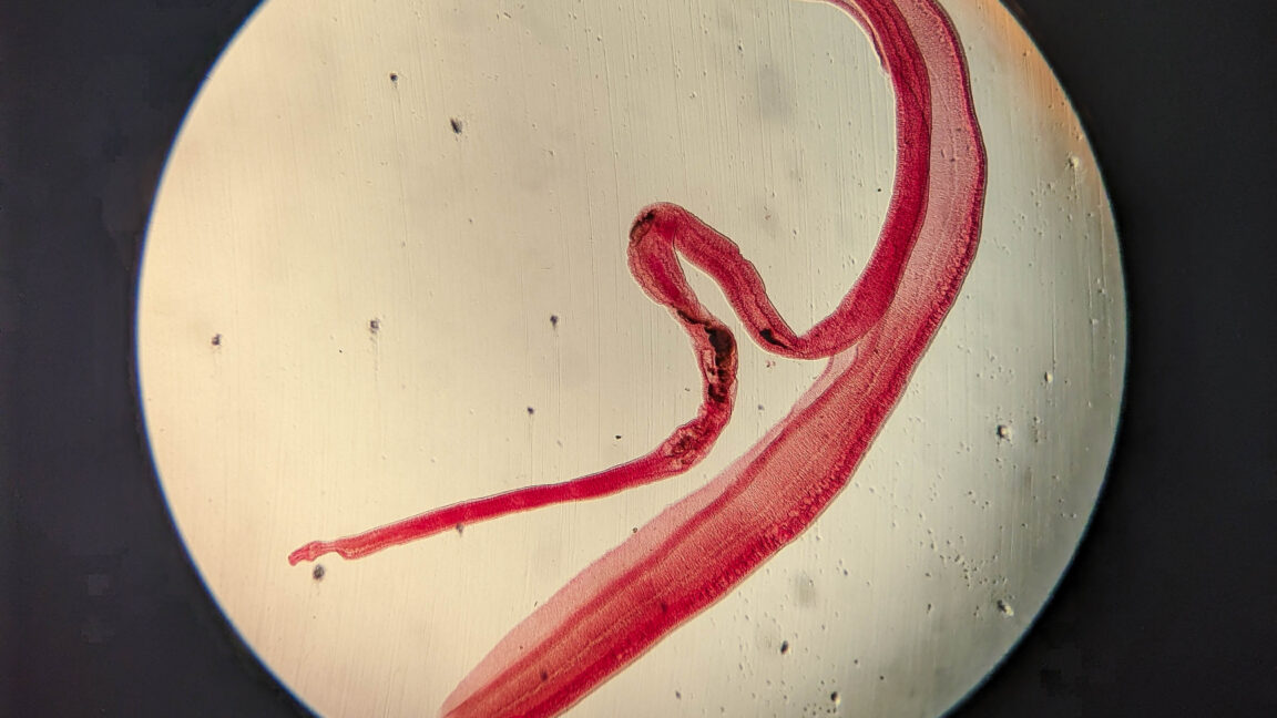
S. japonicum, like its relatives, has a diabolical life cycle including humans. When conditions are right, the eggs hatch into a free-swimming larval form. Late-stage larvae then burst out, grow a forked tail, and swim to find an animal host.
The adult male and female worms live paired together with the female worm tucked into a groove within the thicker male.
Adult worms often go undetected in blood vessels, producing no noticeable symptoms, allowing the infection to become chronic.
The possibility of S. japonicum eggs in the woman’s brain seemed like it could explain her abrupt seizure, so her doctors began diagnostic tests. The woman’s doctors first turned to an old-school lab test called the circumoval precipitin test, which was developed in the 1950s. In this test, doctors get a reference sample of S. japonicum eggs and flood them with a patient’s serum.